IntroductionSchistosomiasis is a chronic neglected tropical parasitic disease that poses a threat to global health. It is caused by parasitic trematode worm (schistosomes or blood flukes) belonging to the class Trematoda, family Schistosomatidae and genus Schistosoma (1). They inhabit the blood stream of homeothermic animals (warm blooded animal). Globally, it was estimated to affects more than two-hundred million populace and it’s regarded as the second public health importance after malaria, causing fatalities of 280,000 people yearly in Africa continent (2; 3).
Profile view of concentrated Asian microbiologist examining sample with help of modern microscope while wrapped up in work at dim laboratoryCharacteristics of Schistosome
They are dioceious, sexually dimorphic; eggs are non operculate with spine found in blood stream of homeothermic animals. There cereariae are furcocercous or brevifurcate in nature (1).
Prevalence and Geographical Distribution
There are several species of schistosome affecting man namely, Schistosoma haematobium causes urinary schistosomiasis, Schistosoma mansoni causes intestinal schistosomiasis, Schistosoma japonicum causes intestinal schistosomiasis, Schistosoma guineensis,Schistosoma intercalatum and Schistosoma mekongi causes intestinal schistosomiasis (3). Globally, an estimated of over 779 million people are at risk from the disease (3). Over 250 million people are infected with Schistosoma spp. worldwide, with 201.5 million of them living in Africa (4; 5).
Schistosoma mansoni is prevalence in Africa, Caribbean and South America while Schistosoma japonicum and Schistosoma haematobium are prevalence in Africa, Eastern Mediterranean and pacific region, the highest prevalence and intensities usually found in school-age children, adolescent and young adults (2). The six Schistosoma spp. that can infect humans have different geographical distributions. S. haematobium has been reported in 54 countries (6) and is the most common species, occurring in sub- Saharan Africa and the Middle East, although a 2013 outbreak of urogenital schistosomiasis was observed (7; 8). S. mansoni is endemic in sub- Saharan Africa, Brazil, the Caribbean islands, Puerto Rico, Suriname and Venezuela and S. japonicum is endemic in the People’s Republic of China and the Philippines, with small foci in Indonesia (9). The remaining species have lower global prevalence with S. guineensis and S. intercalatum is endemic in West Africa and Central Africa and S. Mekongi is prevalence in the southern parts of Cambodi (8; 3).
HIGHLIGHTS ON EPIDEMIOLOGY SUMMARY
Several environmental and socio-economic factors have been identified to be responsible for the continued persistence of this neglected tropical disease (2), they includes;
Presence of natural water bodies which are suitable habitats for the snail intermediate hosts of each parasite, as well as satisfying the needs of man.
Presence of the appropriate snail intermediate hosts of each parasite in the water bodies.
Human contacts with the natural water bodies.
Pollution of water with human urine for haematobium and with excreta for S. mansoni.
Factors which promote the parasites within the snail and it’s transmission to man.
Resistance to infection after the second decade of life and socio-economic status of people (2; 10)
The two common Schistosomiasis are highlighted below
Schistosoma mansoni
This in man causes intestinal schistosomaisis. The worm inhabits the inferior mesenteric vein of man’s intestine. Eggs have lateral spine pointing backward and are non-operculate. The intermidiatehost of the disease is planorbid snails (Biomphalariapheifferri) (10).
Schistosoma haematobium
This worm is responsible for causing urinary schistomiasis in man. It inhabits the vesical plexus that drains the urinary bladder of man. Eggs, which are non-operculate, are provided with terminal spines. In Africa, the intermidiate hosts isaquatic snails of the Bulinus species i.e Bulinus (Physopsis) globosus, Bulinustruncatus, Bulinusforskali, and Bulinusrholfsi (10).
Disease burden
If left untreated, schistosomiasis can result in substantial morbidity and even fatality (3; 2). According to the Global Burden of Disease Study 2016, the global burden of schistosomiasis is estimated at 1.9 million disability- adjusted life years. The morbidity and mortality associated with untreated S. japonicum infection are especially high, probably owing to the relatively higher number of eggs produced by this species than by the others (3).
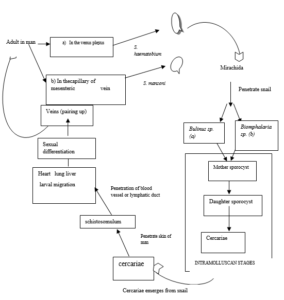
Diagrammatic representation of Life Cycle of S. mansoni and S. Haemetobium Pathophysiology
The progression of schistosome infections can be divided into three general overlapping stages influenced by the duration of the individual’s infection: acute, established active and late chronic infection. These stages differ in egg excretion rates in stool or urine as well as in clinical manifestations and symptoms (3). Organ- specific morbidity can develop during established acute and late chronic stages, caused by the accumulation of parasite eggs and development of fibrosis; the severity of symptoms generally correlates with the intensity of the infection. What organs are affected depends on the infecting Schistosoma spp, as the adult worms of different species localize and lay eggs in specific preferred sites. In addition, serious effects result if adult worms locate and lay eggs in aberrant sites (11).
CLINICAL SYMPTOMS OF SCHISTOSOMIASIS Schistosoma haematobium
The chronic stage is when eggs begin to appear in urine. During this period the following symptoms are observed, they include dysuria, micturation and haematuria. Other pathological effects such as pseudo-tubercle formation, tissue fibrosis, hydroureter, hydronephrosis and uraemia are usually present. All these are due to eggs trapped in the bladder and wall of the ureters (10)
Schistosoma mansoni
Eggs when they are lodged in the mucosa and the sub mucosa of the colon will induce inflammatory reactions such as pseudo-tubercle formation. This condition elicit abscessess which lead to ulceration of the colon. At this stage, symptoms are mainly abdominal pain, diarrhea with blood, mucus and pus. When eggs are carried to the liver and spinal cord, they induce pseudo-tubercle formation and when they are carried to the liver through portal circulation, they give rise to pseudo-tubercle and fibrosis causing liver cirrhosis and portal hypertension (10).
DIAGNOSISClose up view of cells through microscope in modern medicine laboratory
Rectal bladder and liver biopsies may reveal the presence of the eggs or pseudo-tubercle in the tissues.
Immunodiagnostic technique such as intradermal test compliment fixation test (CFT) and the Cercarien Hullen Reaction (CHR)
Demonstration of eggs in urine for haematobium and in stool for S. mansoni.
CONTROL AND TREATMENT
The aim here is to interrupt the live cycle of the parasites so as to cause a break in transmission. This can be achieved by one or a combination of the following:
Reducing human contact with water
Improve sanitation by preventing pollution of water with human urine and excreta
Eradication or reduction of snail population by using mollusicidal chemical e.g. bayluscide.
Ecological modification of water habitats by cutting down marginal vegetation.
Possible use of vaccine to induce immunity.
Chemotherapy can be used to reduce the worm burden or egg production by parasite
References
Bekana, T., Berhe, N., Eguale, T., Aemero, M., Medhin, G., Tulu, B., G/Hiwot, Y., Liang,S., , W And Erko, B. Prevalence and factors associated with intestinal schistosomiasis and human fascioliasis among school children in Amhara Regional State, Ethiopia. Tropical Medicine and Health, (2021) 49:35.
Akinneye, J.O., Fasidi, M.M., Afolabi O.J., and Adesina F.P. (2018) Prevalence of Urinary Schistosomiasis among Secondary School Students in Ifedore Local Government, Ondo State, Nigeria. International Journal of Tropical Diseases, 1:004.
McManus, P. D., David W. Dunne, w. D., Sacko,W, Utzinger, J., Vennervald, J. B. and Nong Zhou, X. Schistosomiasis. Nature Review Diseaese Primers, (2018) 4:13.
Utzinger, J. et al. (2009). Schistosomiasis and neglected tropical diseases: towards integrated and sustainable control and a word of caution. Parasitology 136, 1859.
Hotez, P. J. et al. The Global Burden of Disease Study 2010: interpretation and implications for the neglected tropical diseases. PLoS Negl. Trop. Dis. 8, e2865 (2014).
World Health Organization. Schistosomiasis (WHO, 2017).
Boissier, J. et al. Outbreak of urogenital schistosomiasis in Corsica (France): an epidemiological case study. Lancet Infect. Dis. 16, 971–979 (2016).
Lai, Y.-S. et al. Spatial distribution of schistosomiasis and treatment needs in sub- Saharan Africa: a systematic review and geostatistical analysis. Lancet Infect. Dis. 15, 927–940 (2015).
Colley, D. G., Bustinduy, A. L., Secor, W. E. & King, C. H. Human schistosomiasis. Lancet 383, 2253–2264 (2014).
Dada, E. O., & Jegede, S. O. (2019). Prevalence of Fascioliasis and Dicrocoeliasis in Cattle Slaughtered in Some Abattoirs in Akure Metropolis, Ondo State, Nigeria. International Journal of Pathogen Research, 3(2), 1-7.
Ross, A. G., Vickers, D., Olds, G. R., Shah, S. M. & McManus, D. P. Katayama syndrome. Lancet Infect.7, 218–224 (2007).

 HIGHLIGHTS ON EPIDEMIOLOGY SUMMARY
Several environmental and socio-economic factors have been identified to be responsible for the continued persistence of this neglected tropical disease (2), they includes;
HIGHLIGHTS ON EPIDEMIOLOGY SUMMARY
Several environmental and socio-economic factors have been identified to be responsible for the continued persistence of this neglected tropical disease (2), they includes;
 Diagrammatic representation of Life Cycle of S. mansoni and S. Haemetobium
Pathophysiology
The progression of schistosome infections can be divided into three general overlapping stages influenced by the duration of the individual’s infection: acute, established active and late chronic infection. These stages differ in egg excretion rates in stool or urine as well as in clinical manifestations and symptoms (3). Organ- specific morbidity can develop during established acute and late chronic stages, caused by the accumulation of parasite eggs and development of fibrosis; the severity of symptoms generally correlates with the intensity of the infection. What organs are affected depends on the infecting Schistosoma spp, as the adult worms of different species localize and lay eggs in specific preferred sites. In addition, serious effects result if adult worms locate and lay eggs in aberrant sites (11).
CLINICAL SYMPTOMS OF SCHISTOSOMIASIS
Schistosoma haematobium
The chronic stage is when eggs begin to appear in urine. During this period the following symptoms are observed, they include dysuria, micturation and haematuria. Other pathological effects such as pseudo-tubercle formation, tissue fibrosis, hydroureter, hydronephrosis and uraemia are usually present. All these are due to eggs trapped in the bladder and wall of the ureters (10)
Schistosoma mansoni
Eggs when they are lodged in the mucosa and the sub mucosa of the colon will induce inflammatory reactions such as pseudo-tubercle formation. This condition elicit abscessess which lead to ulceration of the colon. At this stage, symptoms are mainly abdominal pain, diarrhea with blood, mucus and pus. When eggs are carried to the liver and spinal cord, they induce pseudo-tubercle formation and when they are carried to the liver through portal circulation, they give rise to pseudo-tubercle and fibrosis causing liver cirrhosis and portal hypertension (10).
DIAGNOSIS
Diagrammatic representation of Life Cycle of S. mansoni and S. Haemetobium
Pathophysiology
The progression of schistosome infections can be divided into three general overlapping stages influenced by the duration of the individual’s infection: acute, established active and late chronic infection. These stages differ in egg excretion rates in stool or urine as well as in clinical manifestations and symptoms (3). Organ- specific morbidity can develop during established acute and late chronic stages, caused by the accumulation of parasite eggs and development of fibrosis; the severity of symptoms generally correlates with the intensity of the infection. What organs are affected depends on the infecting Schistosoma spp, as the adult worms of different species localize and lay eggs in specific preferred sites. In addition, serious effects result if adult worms locate and lay eggs in aberrant sites (11).
CLINICAL SYMPTOMS OF SCHISTOSOMIASIS
Schistosoma haematobium
The chronic stage is when eggs begin to appear in urine. During this period the following symptoms are observed, they include dysuria, micturation and haematuria. Other pathological effects such as pseudo-tubercle formation, tissue fibrosis, hydroureter, hydronephrosis and uraemia are usually present. All these are due to eggs trapped in the bladder and wall of the ureters (10)
Schistosoma mansoni
Eggs when they are lodged in the mucosa and the sub mucosa of the colon will induce inflammatory reactions such as pseudo-tubercle formation. This condition elicit abscessess which lead to ulceration of the colon. At this stage, symptoms are mainly abdominal pain, diarrhea with blood, mucus and pus. When eggs are carried to the liver and spinal cord, they induce pseudo-tubercle formation and when they are carried to the liver through portal circulation, they give rise to pseudo-tubercle and fibrosis causing liver cirrhosis and portal hypertension (10).
DIAGNOSIS